Revealed

A blog about scientific and medical visualization and all that’s involved.

Archive for July, 2009

Visualizing Lung Anatomy

Posted by Janet on July 27, 2009

One of the projects I’m currently working on at home involves building a 3D model of the right lung. To make sure the model will be as accurate as possible in terms of shape, I would need to know the lung anatomy before I begin building the model.

The first thing I did was going to a library to look at drawings and photographs of lungs. I did a few quick sketches and wrote down some notes on how I will approach this in a 3D program:

sketch1sketch2
Now comes the fun part…

Next, I went to CVS and bought some Crayola modeling clay and began to build a small lung model while looking at online images of the lung. I tried to find as many variations in as many angles as possible, but most resources only showed the standard views. The e-anatomy website very helpful for this initial step, since it provides cross sectional images, labels of structures, and several different ways of presenting the structures. (The website is free, but registration is required to gain full access to the labels and features. High res images and full screen mode are available with a fee, but for our purpose this isn’t necessary.)

The purpose of making a clay model is to get a concrete physical sense of the three-dimensional shape. I find this step very important because it forces you to piece together two-dimensional visuals into a three-dimensional object. It is through this process that I begin to realize the complexity of the shape of a lung.

Below: Clay lung, heart, and a piece of unused clay.

clay-lung

To begin the digital visualization process, I looked further to find visualization tools that would allow me to use actual human data, look at the structures from various angles, and isolate unnecessary structures. Osirix, an open source DICOM viewer, is perfect for the job. It even has data sets available for download. The only problem is…Osirix is Macs only, and I only have access to PCs.

After poking around for a while, I found a similar product called VolView that works on a PC. VolView comes with a $1000 yearly or a $2500 unlimited licencing fee, but it has a 30 day free trial for download. I was able to import a data set from the Osirix website into VolView. My impression of VolView after two days is that it is very easy to navigate, comes with instructions (Help –> Help Topics), gives good results, and has powerful features. One thing I haven’t figured out is whether there is a cut feather that allows the user to trim away unnecessary parts. There seems to be a segmentation tool under the “Analysis” tab, but the instructions for this feature is limited and it is a feature that does not allow “undo.” That is scary. The first time I tried it, it messed up a model I had been playing with for two hours, but it warns you first so you feel like it’s your own fault for not listening. Luckily I was able to reproduce the same result ten minutes later. Here is a screen shot of my lung visualization:

volview-lung
…and a detailed shot of the lung. You can see the bronchi entering the right lung, but notice there are still some artifacts on the side.
volview-lung-detail

*See “Modeling Lung Anatomy” for the next step in this process.

Posted in anatomy, digital 2D/3D, my projects, photography/imaging, techniques | Tagged: , , , , | 4 Comments »

Tours of the Museum Collections

Posted by Janet on July 18, 2009

One of the neat things about being an intern at the Smithsonian is the intern activities. Being 30 minutes away from the main building, we usually can’t attend the lunchtime discussions. The Museum Support Center (MSC) where I work, however, houses part of the museum’s collections so we got some nice tours of the fish and anthropology collection last week.

The first question I asked on each tour was “Can I take pictures?” The answer is yes, but only if it doesn’t go on a website. Images that go on a website have a lot of legal issues that needs to be dealt with, and no one wants to take the time to fill out pages of forms for anyone who wants to post these images. It’s understandable, so I’ll try my best to describe what was seen.

Walking into the fish hall is like walking into a giant freezer. Temperature is kept low to slow the ethanol’s rate of evaporation. Fishes are stored in ethanol-filled jars, with identification labels both on the fish and inside the jars. Special paper and ink are used for these labels so they last, in ethanol, for hundreds of years. This collection reminds me of large library, but with over four million specimens of fish instead of books. Oversized fishes are stored on a separate section, and near the entrance is a shelf that houses the “Oh My” collection. The “Oh My” collection, understandably, contains strange fishes that elicit the “Oh my!” response from people. For example, a two headed shark, a fish that looks like a rock, and a fish that is just too strange to describe. Next, we go to a separate tank room to see the fish that’s even too big for the oversized collection.

The tank room is reminiscent of the bathtub aisle at Costco. On these shelves lie individual metal tanks with sealed lids. The lids can be slid away when they become unlocked, revealing one large fish just under the size of a tank bathing in ethanol. We walked into a preparation room and opened up the tank housing the coelacanth, which was thought to be extinct 65 million years ago until its discovery in 1938.

The museum deals with such a large collection, even after the tours it’s still difficult to comprehend just how big the collection is. The only thing I can think of is that stockroom scene from The Matrix where Neo says “Guns. Lots of guns.” and racks of guns start flying at you. That’s actually a pretty close description to the weapons section of the anthropology collection. The majority of the collection is stored in closed space, but several, including weapons and bones of large mammals, are out in the open due to space constraints. The anthropology collection appears even more spectacular than the fish collection, probably because it was housed in a larger open area rather than separated into a number of smaller rooms. Even so, we went up a total of two flights of stairs and walked through several fire doors before coming back out into the open. Within the collection area, filters run constantly to circulate air flow and filter out dust particulates, and the rows of white cabinets open up to reveal sliding drawers.

Each collection is organized according to the nature of the particular field. This particular collection is organized by geographical region and time. Artifacts included clothing, dolls, baskets, weapons, masks, and much more. Pieces with historical significance are often stored along with related artifacts and copies of historical documents. Even modern pieces such as Korean plastic toys from the 70’s have made their way into the collection. Objects deemed highly sacred by certain cultures are specially cared to make sure only those who are allowed to view or handle them can do so. Like the fish collection, the anthropology collection also accomodates oversized objects. Large objects ranging from figure sized statues to ships sit in custom-built metal frames with fireproof blankets over them. Some frames have wheels to facilitate transportation needs.

This concludes the collection tours. There is no way to open every cabinet in this lifetime. Maybe next week there will be other adventures.

Posted in Smithsonian internship | Tagged: , , , , | Leave a Comment »

Smithsonian Internship

Posted by Janet on July 13, 2009

Third day at the internship, I’m still overwhelmed by everything this place has to offer. Even prior to the start date, the friendliness and efficiency of the staff members really stood out. Weeks ahead of time, I had already received detailed instructions about what to do on the first day. I followed directions diligently like a little kid on a scavenger hunt–“get off the Metro station…walk towards this intersection…go through this particular door, turn right, go through another door and receive a visitor’s pass…” Being nervous about missing the shuttle to the Museum Support Center in Suitland where I’m stationed, I arrived much ahead of time. Staff member Mr. Potter, who just happened to arrive at the same time, kindly introduced me to the cafeteria where employees can enjoy a morning cup of coffee before the museum opens. Mr. Potter then suggested that I kill time by visiting the Sant Ocean Hall. Here’s a glimpse of the exhibit at 8AM, without any visitors:

Smithsonian1

By the third day, I already met nearly 15 other interns. The majority are aspiring young scientists, but there’s a surprising number of illustrators like myself as well. An intern in my section is working her way to apply for a medical illustration program, and I just met another intern who actually goes to the same science illustration program I did four years ago. I’m excited to be surrounded by such a variety of people and all the resources this place has to offer.

Posted in Smithsonian internship | Tagged: , , | Leave a Comment »

The Rest of My Trip In A Nutshell

Posted by Janet on July 8, 2009

Since my last entry, my computer died and I was traveling quite a bit so I lived without the internet for a while. It revived itself a week and half later like nothing happened. No one ever figured out what went wrong, but I spent a week backing everything up “just in case” it dies permanently.

During this period, I traveled for one more week in California visiting various people and attending events, went back to Chicago to pack, and drove to Virginia to settle for my internship at the Smithsonian for the next four months. After meeting lots of artists and scientists and traveling across the country, it’s time to settle down once again to focus on getting my degree.

Today began the first day of my internship, but first, descriptions and images from the time my computer died, and the week before that:

6/1: Aesthetic Prosthetics
Visited “Aesthetic Prosthetics,” an anaplastology clinic located in Pasadena. Spoke to Stefan Knauss, co-owner of the clinic and alumni from the same program I went through at UIC, about materials, techniques, and challenges of opening a business in the field of anaplastology. Mr. Knauss showed me a few prosthetic arms which were quite impressive:
AestheticProsthetics1AestheticProsthetics2

6/5: DreamWorks
Visited the DreamWorks studio in Glendale, CA. It took a while for the visitor’s pass to get approved, but my contact Shannon T. (whom I met through this blog) and his coworkers eventually got me in. I had a nice chat with Shannon before he took me on a tour around the studio to see the flow of the production process.

6/7-6/8: visiting Ed Ross and Sandy Ross
I flew to San Francisco and took a ferry to Marin County before meeting up with entomologist Ed Ross and his wife Sandy Ross. Dr. Ross is now 94 years old and still actively publishing. Sandy made goose eggs for breakfast. Notice the size of this yolk!
GooseEgg

6/10: Stanford Lab and phone interview with Anatomical Travelogue
On this date I visited Dr. Paul Brown’s at Stanford University, who took me on a special tour of the medical campus and gave me an in-depth look at the medical visualization works taking place at Stanford. Special thanks to Sarah H., one of Dr. Brown’s interns and my former classmate, for giving me a detailed look at the current works.

This morning I also had a phone interview with Anatomical Travelogue, a medical 3D visualization company based in New York. Unfortunately there was a major schedule conflict due to my prior commitment to the Smithsonian, so in the end it didn’t work out :-/

6/12: California Academy of Sciences and de Young Museum
On this gloomy San Francisco day, I went with a friend to check out the relatively new Cal Academy building and the nearby de Young Museum. Here I am, standing on top of the “living roof” of the Cal Academy of Sciences.
CalAcademyRoof

6/16: Molecular Graphics Lab
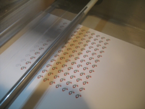
Drove two and half hours from Pasadena to La Jolla to visit the Molecular Graphics Lab. I found out about this lab through the 2008 AMI conference when lab director Dr. Arthur Olson and graduate student/medical illustrator Graham Johnson lectured on the benefits of using physical molecular models as research or educational tools. Dr. Olson, Graham Johnson, and other professors and students I met in the lab showed me different processes involved in making both digital and physical molecular models.
MGL1Above: Rapid prototyping machine printing an array of molecules.
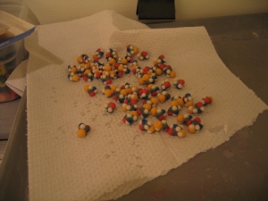
Below: The finished molecules.

MGL2MGL3Above: A display case filled with molecular models.
Below: Comparing relative sizes of molecules.

MGL4

6/19: Huntington Library, corpse flower blooming
When news of the blooming corpse flower reached us, we decided to visit and smell the odor. A day too late, the flower already wilted and the smell had reduced from the powerful “rotten meat” to a weak “rotten vegetable” smell.
CorpseFlower

6/20-6/21: IMATS (International Make-Up Artist Trade Show)
First thing at the show, I give prosthetic make-up a try.
Below: applying spirit gum to skin

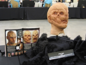
IMATS1Below: Bullet hole on skin with fake blood. Move one more foot away and you won’t see the edges anymore.IMATS2Below: Airbrush demoIMATS3Below: Museum area showing masks and props from movies.IMATS4Below: Student competition (second day, fantasy make-up)IMATS5
Posted in places, Uncategorized | 1 Comment »